

Surgical training has always relied on human tissue to teach what no textbook can. How freshly-frozen cadavers improve surgical skills training comes down to one simple fact: the tissue behaves like a living patient, not a preserved museum piece. Freshly-frozen cadavers are donor bodies conserved through rapid sub-zero freezing rather than chemical embalming. That single difference in method changes everything about how the specimen feels under a scalpel.
Because there's no formalin soaking into the cells, the tissue keeps its natural pliability. Joints bend, fascia slides, muscle separates the way it would in an operating room. This is why donor-based education built around fresh-frozen specimens has become the standard for programs that want trainees to walk into surgery with real hands-on surgical education already under their belt, not just theoretical knowledge from a lecture hall.
Why do programs prefer freshly-frozen tissue over embalmed alternatives?
Formalin-based preservation has a long history, but it comes with a tradeoff. Embalmed tissue turns stiff, grayish, and rubbery. Tactile feedback that surgeons depend on, the give of a vessel wall, the resistance of a ligament, gets flattened out. Tissue handling on an embalmed specimen feels nothing like the real thing, and residents who train exclusively on embalmed models often need extra time to adjust once they reach live procedures.
Fresh-frozen specimens skip that distortion entirely. Joints keep their range of motion, sliding tissue planes stay intact, and cutting resistance stays close to what a surgeon meets in the OR. A review published in Anatomical Sciences Education lays out how modern preservation methods affect anatomical realism, and the conclusion tracks with what most lab directors already know from experience: for advanced clinical skills development, human tissue realism matters more than convenience.
How do freshly-frozen models compare to virtual simulations?
Screen-based simulators have a real place in surgical education. They're fast, repeatable, and don't require a lab full of biohazard protocols. But framing this as a competition misses the point, surgical simulation and cadaveric training solve different problems, and a well-built curriculum uses both.
The benefits of computerized operative simulation
Operative simulation software is good at building early hand-eye coordination. Trainees can repeat a suturing motion a hundred times in an afternoon, track their speed, and see exactly where their instrument path drifted. It's a low-stakes way to build muscle memory before ever touching a scalpel to skin.
The irreplaceable value of real tissue handling
What a simulator can't do is replicate the moment a suture tears through weakened tissue, or the way fat layers differ in thickness from one patient to the next. Procedural confidence comes from encountering those surprises on a real specimen, not from a screen that resets with a click.
What anatomical benefits make fresh tissue training highly realistic?
The realism isn't just about texture. It's about spatial relationships that stay true to life, where a nerve actually runs relative to a vessel, how dense connective tissue really is layer by layer. No two donors are identical, and that anatomical variation is exactly what prepares residents for the unpredictability of a real patient on the table.
Mimicking real-world laparoscopic skills training
Inside a closed cavity, angle limitations and tool feedback become the whole game. Laparoscopic skills training on fresh-frozen tissue forces trainees to work with the same visualization constraints they'll face in a live procedure, something a flat screen simply can't reproduce.
Fostering clinical skills development and tissue manipulation
Suturing, cutting, and blunt dissection all feel different when practiced on tissue that hasn't been chemically hardened. A study in PMC found construct validity for fresh-frozen cadavers in minimal access surgery training, showing measurable gains in surgical competency after repeated laparoscopic practice on real specimens.
Which surgical specialties benefit most from cadaveric workshops?
Not every specialty needs the same depth of tissue realism, but a few rely on it heavily. Orthopedics, neurosurgery, ENT, and plastic surgery all involve deep anatomical corridors and complex reconstructions where a wrong turn has real consequences. A cadaver workshop built around joint replacements, sinus dissections, or vascular shunt practice gives residents a rehearsal space that mirrors the operating room far more closely than any other training method.
Orthopedic and neurosurgical deep-access training
Navigating around bone, nerve bundles, and major vascular networks demands tissue that responds like the real thing. Pliable, fresh-frozen specimens let trainees practice the same careful navigation they'll need when the margin for error is measured in millimeters.
Advanced soft-tissue reconstruction and flap harvesting
Plastic and reconstructive specialties depend on realistic tissue planes to practice microvascular connections and flap transfers. A systematic review in PMC looked at cadaveric workshops across postgraduate surgical training and found consistent gains in technical skill and trainee confidence, reinforcing why specialty procedure training so often centers on this kind of preclinical procedural practice and advanced surgical rehearsal.
What are the limitations and operational challenges of frozen specimens?
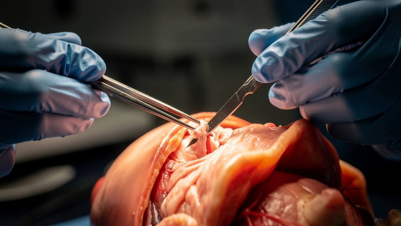
Fresh-frozen tissue isn't without drawbacks. Running a cadaver lab training program takes planning that goes well beyond booking a room and inviting residents.
Logistical and preparation hurdles
Specimens need a controlled thaw window, too early and the tissue starts to degrade, too late and the lab schedule falls apart. Biohazard protocols, sub-zero storage, and strict handling rules all add layers of preparation that an R&D cadaver lab has to manage before a single incision is made.
The lack of functional physiologic feedback
Without a perfusion pump, a fresh-frozen specimen has a bloodless field. There's no active bleeding, no tissue healing response, no way to simulate a hemorrhage in real time. For medical device testing that depends on physiologic reaction, this is a real constraint, and it's one reason some labs pair cadaveric sessions with live-tissue or simulation models to cover what frozen specimens can't.
Additional independent research comparing long-term outcomes across training methods would help clarify how much of the skill gain from cadaveric practice carries directly into operating room performance.
| Training Method | Tactile Realism | Best Use Case |
|---|---|---|
| Fresh-frozen cadaver | High | Advanced procedural rehearsal, specialty workshops |
| Embalmed cadaver | Low to moderate | Basic anatomy orientation |
| Virtual simulator | None | Early coordination and speed metrics |
Is fresh-frozen cadaver training safe for repeated use?
Yes, within proper cold-chain handling. Specimens are re-frozen between sessions when protocols allow, though repeated thaw cycles do accelerate tissue degradation over time.
How long can a thawed specimen be used before it degrades?
Most labs work within a window of a few days, depending on storage temperature and how the specimen was initially prepared before freezing.
Can virtual simulators fully replace cadaver labs?
No. Simulators build coordination and speed, but they can't reproduce the variable resistance and anatomical variation found in real human tissue.
Which specialties rely most on cadaveric workshops?
Orthopedics, neurosurgery, ENT, and plastic surgery lean on cadaveric training the most, given the anatomical precision each field demands.
About the Business
Biotech Anatomy LTD runs a modern facility in Israel built around practical anatomy and surgical education, supporting surgeons, medical students, healthcare professionals, researchers, and medical device developers who need real donor-based training environments. The team handles the procurement and preparation side so that residency programs and device companies can focus purely on the work itself, and for anyone organizing a session, their cadaver lab anatomy training setup covers everything from specimen readiness to full lab support. Biotechanatomy positions itself as a working partner for R&D and education alike, not just a facility rental.